A common and dangerous misconception about heart attacks is that they result from a plaque gradually growing large enough to completely block a coronary artery—like a pipe slowly clogging. Clinical pathology data reveals a very different picture: in approximately 75% of acute myocardial infarctions, the obstructing lesion was not the largest plaque in the vessel.
The Clot, Not the Plaque, Kills
A common and dangerous misconception about heart attacks is that they result from a plaque gradually growing large enough to completely block a coronary artery - like a pipe slowly clogging. Clinical pathology data reveals a very different picture: in approximately 75% of acute myocardial infarctions, the obstructing lesion was not the largest plaque in the vessel.
The actual mechanism is more acute and more tractable: a vulnerable plaque - often only mildly obstructive - develops a fissure or ruptures, exposing its lipid-rich, collagen-rich core to flowing blood. The blood responds to this exposed tissue by initiating the clotting response. A thrombus (blood clot) forms rapidly on top of the plaque, and within minutes, a vessel that was 40% occluded becomes completely blocked.
The pathological event that kills is platelet hyperactivation and pathological thrombus formation - not the plaque itself. Understanding this mechanism reveals why anti-thrombotic nutrition is as clinically important as lipid management.
Platelet Biology: From Sentinels to Killers
Platelets (thrombocytes) are small, anucleate blood cells derived from megakaryocytes in the bone marrow. Their primary physiological role is hemostasis - stopping bleeding when vascular integrity is breached. They achieve this through a tightly regulated sequence of activation, adhesion, and aggregation.Under pathological conditions - particularly in the context of atherosclerosis, oxidative stress, and systemic inflammation - platelets become hyperactivated: abnormally responsive to activation signals, aggregating more readily, and forming more robust clots than the physiological situation demands.
The Three Stages of Pathological Platelet Activation
Stage 1: AdhesionWhen the endothelium is damaged or plaque ruptures, subendothelial collagen and von Willebrand Factor (vWF) are exposed. Platelets express Glycoprotein VI (GPVI) receptors that bind collagen directly, and GPIb-IX-V receptors that bind vWF. This initial adhesion anchors platelets to the injury site.
Stage 2: ActivationAdhered platelets undergo a conformational change - from discoid to spiky, amoeba-like forms - and release the contents of their internal granules:
- Dense granules: Release ADP (the most potent platelet activator), ATP, serotonin, and calcium
- Alpha granules: Release fibrinogen, vWF, P-selectin, and platelet factor 4
ADP activates two receptors on neighboring platelets: P2Y1 and P2Y12 (the target of clopidogrel). P2Y12 activation inhibits adenylyl cyclase → reduces cAMP → promotes platelet activation.
Simultaneously, activated platelets convert arachidonic acid (from membrane phospholipids) via cyclooxygenase-1 (COX-1) to Thromboxane A2 (TXA2) - a potent vasoconstrictor and platelet activator that binds TP receptors and amplifies aggregation.
Stage 3: AggregationPlatelet activation triggers a conformational change in surface Glycoprotein IIb/IIIa (GPIIb/IIIa) integrin receptors. In their resting state, GPIIb/IIIa cannot bind fibrinogen. After activation, they transition to a high-affinity state and bind fibrinogen with high avidity.
Since fibrinogen has two ApoB binding sites, it cross-links adjacent platelets, creating platelet aggregates that grow exponentially as each activated platelet recruits more. The primary platelet plug forms within seconds.
The Coagulation Cascade: Converting the Plug to a Clot
The primary platelet plug is fragile. To create a stable, durable thrombus, the coagulation cascade is simultaneously activated, ultimately producing fibrin from fibrinogen - a fibrous protein that reinforces the platelet plug into a mechanically stable clot.
The Two Pathways
Extrinsic Pathway (primary pathway in plaque rupture):- Ruptured plaque exposes Tissue Factor (TF) → binds Factor VIIa
- TF-VIIa complex activates Factor X and Factor IX
- Factor Xa assembles the prothrombinase complex → converts prothrombin (Factor II) to thrombin (Factor IIa)
- Exposed subendothelial collagen activates Factor XII
- Cascade amplifies through Factors XI → IX → VIII → X
- Also converges on prothrombinase complex → thrombin
- Thrombin (Factor IIa) is the master enzyme of coagulation
- Converts soluble fibrinogen to fibrin monomers
- Activates Factor XIII, which crosslinks fibrin monomers into a mechanically stable fibrin mesh
- Simultaneously activates more platelets (via PAR-1 receptor) → amplifies aggregation
The result: a mechanically robust thrombus of fibrin-enmeshed, aggregated platelets - capable of completely occluding a coronary artery within minutes.
What Creates Platelet Hyperactivation?
In healthy individuals with intact endothelium, counter-regulatory mechanisms prevent pathological platelet activation:
- Prostacyclin (PGI2): Produced by endothelial cells, it activates adenylyl cyclase in platelets → raises cAMP → powerful platelet inhibitor
- Nitric Oxide (NO): Also produced by endothelial cells; activates guanylyl cyclase → raises cGMP → inhibits platelet activation
- CD39/NTPDase1: Expressed on endothelial cells; degrades the ADP that activates P2Y receptors
In the setting of endothelial dysfunction - the state in which atherosclerosis develops - these inhibitory systems fail:
- PGI2 production decreases
- NO bioavailability falls (consumed by oxidative stress)
- CD39 expression declines
The platelet-endothelium interaction tips from inhibition to activation. Platelets become hyperresponsive.
Additional drivers of platelet hyperactivation include:
| Driver | Mechanism |
|---|---|
| Oxidized LDL | Directly activates platelets via CD36 receptor; depletes NO |
| Elevated Lp(a) | Anti-fibrinolytic effect; competitive plasminogen displacement |
| Elevated fibrinogen | Increased substrate for clot formation |
| Elevated homocysteine | Promotes thrombomodulin suppression; increases procoagulant factors |
| Diabetes/Hyperglycemia | Glycation of platelet surface proteins increases reactivity |
| Systemic inflammation | TNF-α upregulates TF expression; IL-6 stimulates fibrinogen production |
| Omega-6 excess | Arachidonic acid → TXA2 amplification |
The TXA2/PGI2 Balance: The Central Thrombotic Dial
The ratio of Thromboxane A2 (TXA2) to Prostacyclin (PGI2) is perhaps the most important determinant of platelet aggregation tendency in vivo:
- TXA2 (from platelets): Potent vasoconstrictor + platelet activator
- PGI2 (from endothelium): Potent vasodilator + platelet inhibitor
In healthy vessels: PGI2 dominates → anti-thrombotic balance
In atherosclerotic, dysfunctional endothelium: TXA2 dominates → prothrombotic state
Aspirin's mechanism: Irreversibly acetylates COX-1 in platelets, blocking TXA2 synthesis. Because platelets lack nuclei and cannot regenerate COX-1, this effect lasts the platelet's 7-10 day lifespan. This is why low-dose aspirin is anti-thrombotic.Natural Anti-Thrombotic Strategies
Several nutritional compounds modulate the TXA2/PGI2 balance and platelet activation through distinct mechanisms:
Omega-3 Fatty Acids (EPA/DHA)
- EPA competes with arachidonic acid as COX-1/2 substrate, producing TXA3 (weak aggregator) instead of TXA2 (strong aggregator)
- DHA reduces GPIIb/IIIa expression on platelet membranes, reducing aggregation tendency
- Omega-3 supplementation reduces platelet aggregation by 25-45% in clinical studies
Nitric Oxide Precursors (L-Arginine, L-Citrulline)
- Increasing NO bioavailability raises platelet cGMP → directly inhibits GPIIb/IIIa activation
- Restores the endothelial PGI2/NO anti-thrombotic environment
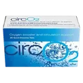
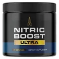
- CircO2 and Nitric Boost Ultra target this pathway directly through sublingual nitric oxide precursor delivery
Nattokinase
- A serine protease derived from natto (fermented soy)
- Directly cleaves fibrin, dissolving existing thrombus
- Inhibits PAI-1 (Plasminogen Activator Inhibitor-1), restoring the fibrinolytic system
- Inhibits platelet aggregation independently of fibrinolysis
Garlic (Allicin/Ajoene)
- Ajoene inhibits fibrinogen binding to GPIIb/IIIa receptors
- Allicin inhibits TXA2 synthesis
- Clinical data shows 40% reduction in platelet aggregation with therapeutic garlic extract
Curcumin
- Inhibits TXA2 receptor (TP receptor) signaling
- Reduces platelet P-selectin expression
- Decreases fibrinogen levels
Conclusion
The fatal event in most myocardial infarctions is not a plaque - it is a thrombus. The entire sequence from plaque rupture to arterial occlusion unfolds within minutes, driven by platelet hyperactivation, TXA2 amplification, and the coagulation cascade converting fibrinogen into fibrin mesh.
Platelet hyperactivation is not random - it is the predictable consequence of endothelial dysfunction, elevated oxidized LDL, elevated Lp(a), and the systemic inflammation that characterizes metabolic syndrome. Addressing the root causes of platelet hyperresponsiveness - restoring the PGI2/NO anti-thrombotic endothelial environment, correcting the omega-6/omega-3 imbalance, and supporting fibrinolytic capacity - is as essential to cardiovascular protection as managing lipid levels.
For the endothelial root cause that enables thrombogenesis, see: how-endothelial-dysfunction-silently-damages-your-cardiovascular-system. For the Lp(a) anti-fibrinolytic contribution, see: lipoprotein-a-residual-cardiovascular-risk-atherogenesis.
Scientific References & Validation
Full citations with PMID links, methodology notes & evidence ratings on puresupphub.com